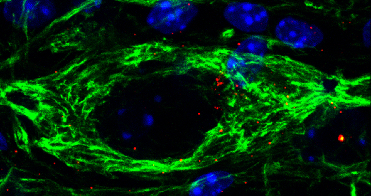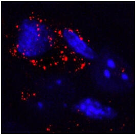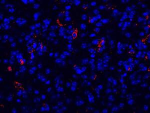
Fig. 1 - High magnification of immunofluorescence staining showing autophagic vesicles (LC3-positive, red) in SMI32-positive motoneuron (green) located in the ventral horn of the SMA lumbar spinal cord. Nuclear staining (DAPI, blue).
Cell Death and Disease, 20 December 2017
Inhibition of autophagy delays motoneuron degeneration and extends lifespan in a mouse model of spinal muscular atrophy
Antonio Piras 1,2,3 , Lorenzo Schiaffino 1,2, , Marina Boido 1,2 , Valeria Valsecchi 1,2 , Michela Guglielmotto 1,2 , Elena De Amicis 1,2 , Julien Puyal 4 , Ana Garcera 5 , Elena Tamagno 1,2 , Rosa M Soler 5 & Alessandro Vercelli 1,2
Spinal muscular atrophy (SMA) is a recessive autosomal neuromuscular disease, due to homozygous mutations or deletions in the telomeric survival motoneuron gene 1 (SMN1). SMA is characterized by motor impairment, muscle atrophy, and premature death following motor neuron (MN) degeneration.
Emerging evidence suggests that dysregulation of autophagy contributes to MN degeneration. We here investigated the role of autophagy in the SMNdelta7 mouse model of SMA II (intermediate form of the disease) which leads to motor impairment by postnatal day 5 (P5) and to death by P13.
We first showed by immunoblots that Beclin 1 and LC3-II expression levels increased in the lumbar spinal cord of the SMA pups. Electron microscopy and immunofluorescence studies confirmed that autophagic markers were enhanced in the ventral horn of SMA pups. To clarify the role of autophagy, we administered intracerebroventricularly (at P3) either an autophagy inhibitor (3-methyladenine, 3-MA), or an autophagy inducer (rapamycin) in SMA pups.
Motor behavior was assessed daily with different tests: tail suspension, righting reflex, and hindlimb suspension tests. 3-MA significantly improved motor performance, extended the lifespan, and delayed MN death in lumbar spinal cord (10372.36 ± 2716 MNs) compared to control-group (5148.38 ± 94 MNs).
Inhibition of autophagy by 3-MA suppressed autophagosome formation, reduced the apoptotic activation (cleaved caspase-3 and Bcl2) and the appearance of terminal deoxynucleotidyl transferase dUTP nick end labeling (TUNEL)-positive neurons, underlining that apoptosis and autophagy pathways are intricately intertwined. Therefore, autophagy is likely involved in MN death in SMA II, suggesting that it might represent a promising target for delaying the progression of SMA in humans as well.
>> read full article


