The steep increase in the microscopy techniques, as well as their complexity in terms of sample preparation and analysis, calls for a centralisation of resources, in order to users to have one reference center where several expertise are available. PICO (Platform for Imaging at Cavalieri Ottolenghi) is located at the Neuroscience Institute Cavalieri Ottolenghi.
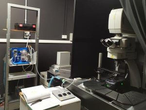
The Facility is equipped with excellent quality light microscopes , in particular, two confocal microscopes (Leica SP5 and Nikon) a Nikon ViCo system , and a high throughput Zeiss Axioscan image acquisition system. Moreover, a two-photon microscope Nikon (A1MP) and Light Sheet microscope (LaVision) have been recently installed.
The facility is also equipped for electron microscopy preparation and imaging, with one TEM available. There are also several imaging systems equipped for high-resolution morphometric investigations, densitometry quantitative autoradiography, as well as workstations for image processing and statistical analysis. Two Neurolucida systems are included in the microscopy facility. Sliding or rotational microtomes, 3 vibratomes and 4 cryostats are available for neuro-histological studies.
Tools
Scanning Electron Microscope (SEM) Zeiss Gemini 460
Equipped with:
STEM Module
High-resolution Volume BSE detector
Zeiss “Volutome” automated serial section and imaging system with focal CC compensator

Leica TCS SP5 Confocal Microscope
Microscope: Leica DM6000CS
High-efficiency detection SP AOBS (Acousto-Optical Beam Splitter)
Motorized stage
Lasers: Argon, 65 mW, 488 nm – HeNe, 1mW; 543 nm – HeNe, 10 mW, 633 nm
UV: Diode, 50 mW, 405 nm
Objectives: 20x/0.50 (dry), 40x/1.25 (oil), 63x/1.40 (oil)
Dichroic: RT 30/70; Substrates; TD 488/543/633; DD 458/514; RSP 500; DD 488/543
Computer: HP xw8400 workstation with Intel Xeon 5160 CPU, 2 GB RAM
Windows XP Professional
Leica LAS AF 2.6.0.7266
Simultaneous 4-channel acquisition


Confocal Microscope NIKON C1
Eclipse digital photo-microscope
Spectral detection unit
Lasers: 488nm Multiline Argon, He Ne 543nm, He Ne 640nm, Violet diode 408nm
Objectives: 20X, 40X, 62X
Simultaneous acquisition on 3 channels + 4th separate channel
Filters: DAPI, Green (Cy2, Alexa488), Red (Cy3)
Imaging Software: Neurolucida and Neurolucida Explorer (11.03 ver.), Stereoinvestigator (11.03 ver.)

Microscope 2-photon
Nikon High-speed multi-photon confocal microscope A1RMP
Motorized stage with adapters for in vivo, ex-vivo and in vitro imaging
Scanning system: galvano scanner and resonant scanner for high speed acquisition
Femtosecond pulsed IR-laser (Ti:Sapphire laser system; Coherent) tunable from 700 nm to 1080 nm.
Objectives: CFI60 Planfluor 10x A.N.0,3 d.l. 16 mm; CFI75 LWD 16xW NIR A.N.0,80 d.l. 3,0 mm; CFI75 LWD Apo 25xW A.N.1 d.l. 2,0 mm; CFI60 Apo 40xW NIR A.N.0,80 d.l. 3,5 mm
Detector 4 Ch GaAsP NDD high sensitivity detector


Light Sheet Microscope
Andor Zyla 5.5 sCMOS Camera
Infinity Corrected Optics Setup
Lenses: 4X and 12X (organic solvent dipping objective lens)
Laser: 488-85, 561-100, 639-70
Workstation for the management and analysis of 3D images, by IMARIS software

Zeiss Axioscan Z.1
Illuminator: Colibri 7, R[G/Y]CBV-UV
Camera Sets Hitachi HV-F203 and Orca Flash 4.0 V3
Objectives: ‘Fluar’ 5x/0.25 M27, ‘Plan-Apochromat’ 10x/0.45 M27, ‘Plan-Apochromat’ 20x/0.8 M27, ‘Plan-Apochromat’ 40x/0.95 M27
Filters: 56/90/91/92 HE LED, filter set 108 HE LED, 38 HE eGFP without shift, 43 HE Cy3 without shift, 96 HE
25 Trays x4 slides 26x76mm; 5 Trays x2 slides 52x76mm; 1 Tray x1 slide 102x76mm
Computer: Intel Xeon Gold Processor 6134 (hp Z6) Premium Zeiss 60A Workstation; No. 6 Memories 32GB (1×32) DDR4-2666 (hp Z6); NVIDIA Quadro P4000 8GB Graphic Card; Zen 2.6 slidescan hardware
Workstation: High End ZEISS 55A R2; Zen 2.6 desk Hardware; No. 4 Memories 32GB (2×16) DDR4-2133 (Z840); NVIDIA Quadro M6000 24GB D Video Card

Neurolucida (workstation 1)
Nikon Eclipse E600 Microscope
Motorized stage
Microfire Camera 2-Megapixel Color Imaging (1600×1200)
Lenses: 2X-4X-10X-20X-40X-100X
Filters: DAPI, Green (Cy2, Alexa488), Red (Cy3)
Imaging Software: Neurolucida and Neurolucida Explorer (11.03 ver.), Stereoinvestigator (11.03 ver.)

Neurolucida (workstation 2)
Microscope Nikon Eclipse 80i
Color digital camera 3/4″ CCD chip color, 1.92 MP, 1600H x 1200V pixels
Motorized stage
Microfire Camera 2-Megapixel Color Imaging (1600×1200)
Lenses: 4X-10X-20X-40X-60X-100Xoil
Filters: DAPI, Green (Cy2, Alexa488), Red (Cy3), dark red
Imaging Software: Neurolucida and Neurolucida Explorer
Transmission Electron Microscope (TEM)
JEOL JEM-1010
 Electron Source: Tungsten filament
Accelerating Voltage 40kv-100kv
Magnification Standard Mag Mode 600x to 500,000x
Users
- NICO research groups
- Departments of the University of Turin
- companies and private research centres with whom the members of the Departments of the University of Turin have ongoing collaborations involving the use of imaging analysis
- companies and private research centres outside the University of Turin
Service delivery
The use of the equipment is subject to a reservation to be made online after being accredited as internal or external users, using the management software BookMyLab
Management Committee
Silvia De Marchis, Marina Boido, Annalisa Buffo
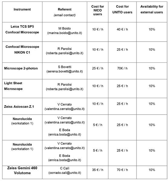
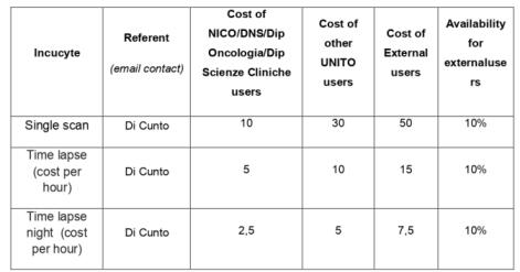
Service costs
The costs of use are calculated annually in order to monitor the use of the instruments.
Price List


Internal and external users are trained in the use of the instruments by the referents.
E Boda enrica.boda@unito.it
M Boido marina.boido@unito.it
V Cerrato valentina.cerrato@unito.it
R Parolisi roberta.parolisi@unito.it
F Di Cunto ferdinando.dicunto@unito.it
