International Journal of Molecular Sciences , 12 March 2021
A First Phenotypic and Functional Characterization of Placental Extracellular Vesicles from Women with Multiple Sclerosis
Martire S 1,2,** , Montarolo F 1,2,3** , Spadaro M 1,2, , Perga S 1,2,4 , Sforza M.L 1,2 , Marozio L 5 , Frezet F 5 , Bruno S 6 , Chiabotto G 6 , Deregibus M.C 7 , Camussi G 6 , Botta G 8 , Benedetto C 5 , Bertolotto A 1,2
Pregnancy is a unique situation of physiological immunomodulation, as well as a strong Multiple Sclerosis (MS) disease modulator whose mechanisms are still unclear. Both maternal (decidua) and fetal (trophoblast) placental cells secrete extracellular vesicles (EVs), which are known to mediate cellular communication and modulate the maternal immune response.
Their contribution to the MS disease course during pregnancy, however, is unexplored. Here, we provide a first phenotypic and functional characterization of EVs isolated from cultures of term placenta samples of women with MS, differentiating between decidua and trophoblast. In particular, we analyzed the expression profile of 37 surface proteins and tested the functional role of placental EVs on mono-cultures of CD14+ monocytes and co-cultures of CD4+ T and regulatory T (Treg) cells.
Results indicated that placental EVs are enriched for surface markers typical of stem/progenitor cells, and that conditioning with EVs from samples of women with MS is associated to a moderate decrease in the expression of proinflammatory cytokines by activated monocytes and in the proliferation rate of activated T cells co-cultured with Tregs. Overall, our findings suggest an immunomodulatory potential of placental EVs from women with MS and set the stage for a promising research field aiming at elucidating their role in MS remission.
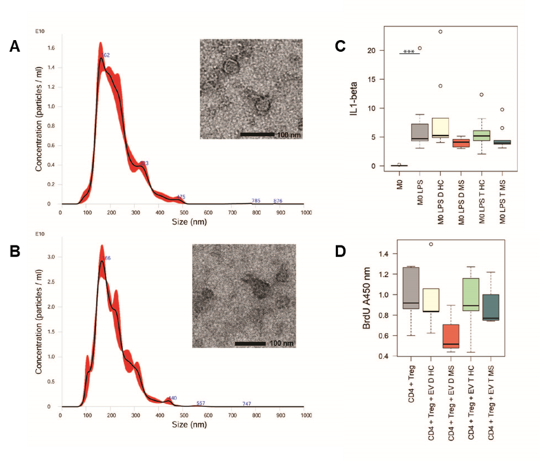
Panel A and B shows shape and size of placental extracellular vesicles of a healthy woman (A) and a woman with MS (B), obtained by transmission electron microscopy and nanoparticle tracking analysis. Panel C shows the expression level of the pro-inflammatory cytokine IL1-β in cell cultures of untreated monocytes (M0), monocytes activated with pro-inflammatory stimuli (M0 LPS) and treated with extracellular vesicles secreted from decidua and trophoblast tissues of healthy women and women with MS. Panel D shows the proliferation level of CD4 + T lymphocytes co-cultured with regulatory T cells untreated (CD4 + Treg) and treated with extracellular vesicles secreted from decidua and trophoblast tissues of healthy women and women with MS.
1
Neuroscience Institute Cavalieri Ottolenghi (NICO), Orbassano, 10043 Turin, Italy
2
Neurology-CRESM (Regional Reference Center for Multiple Sclerosis), AOU San Luigi Gonzaga, Orbassano, 10043 Turin, Italy
3
Department of Molecular Biotechnologies and Health Sciences, University of Turin, 10124 Turin, Italy
4
Department of Neuroscience Rita Levi-Montalcini, University of Turin, 10124 Turin, Italy
5
Department of Surgical Sciences, Obstetrics and Gynaecology, University of Turin, 10124 Turin, Italy
6
Department of Medical Sciences and Molecular Biotechnology Center, University of Turin, 10124 Turin, Italy
7
2i3T Business Incubator and Technology Transfer, University of Turin, 10124 Turin, Italy
8
Department of Pathology, Città della Salute e della Scienza di Torino, 10124 Turin, Italy
**T hese authors contributed equally to this work.


