Acta Neuropathologica Communications , 18 January 2023
Sleep fragmentation affects glymphatic system through the different expression of AQP4 in wild type and 5xFAD mouse models
Vasciaveo V, Iadarola A, Casile A, Dante D, Morello G, Minotta L, Tamagno E, Cicolin A, Guglielmotto M.
Research Group NICO: Aging and Alzheimer's Disease
Alzheimer’s disease (AD) is characterized by genetic and multifactorial risk factors. Many studies correlate AD to sleep disorders. In this study, we performed and validated a mouse model of AD and sleep fragmentation, which properly mimics a real condition of intermittent awakening. We noticed that sleep fragmentation induces a general acceleration of AD progression in 5xFAD mice, while in wild type mice it affects cognitive behaviors in particular learning and memory. Both these events may be correlated to aquaporin-4 (AQP4) modulation, a crucial player of the glymphatic system activity.
In particular, sleep fragmentation differentially affects aquaporin-4 channel (AQP4) expression according to the stage of the disease, with an up-regulation in younger animals, while such change cannot be detected in older ones. Moreover, in wild type mice sleep fragmentation affects cognitive behaviors, in particular learning and memory, by compromising the glymphatic system through the decrease of AQP4. Nevertheless, an in-depth study is needed to better understand the mechanism by which AQP4 is modulated and whether it could be considered a risk factor for the disease development in wild type mice.
If our hypotheses are going to be confirmed, AQP4 modulation may represent the convergence point between AD and sleep disorder pathogenic mechanisms.
Open Access - read more
Our researchers. From the left: Valeria Vasciaveo, Elena Tamagno, Giulia Morello e Michela Guglielmotto
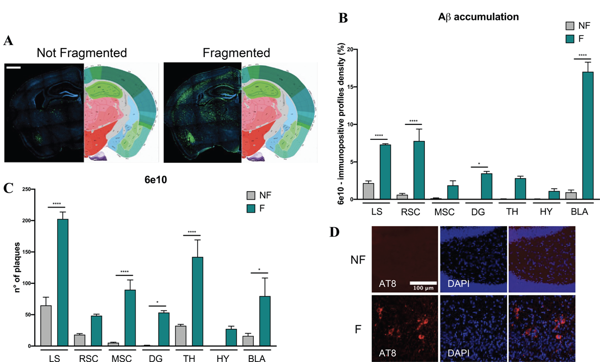
A Representative images of immunohistochemistry acquired with Axioscan microscope. Scale bar 1000 μm. Aβ plaques are labeled in green and the nuclei with DAPI in blue. B Histogram of Aβ plaques analyzed through the percentage of pixels, after the same threshold is set for all the region of interests. C Histogram of Aβ plaque number. D Representative images of immunohistochemistry of AT8. NF not fragmented mice, F fragmented mice, LS lateral septum, RSC retrosplenial cortex, MSC motor-sensory cortex, DG dentate gyrus, TH thalamus, HY hypothalamus, BLA basolateral amygdala. The data are mean standard error of the mean (SEM). Each data point represents an individual animal. * p < 0.05; **** p < 0.0001 versus control by one-way ANOVA followed by Bonferroni post-hoc test, n = 4 per condition


