Experimental Neurology , August 2016
Successful in vivo MRI tracking of MSCs labeled with Gadoteridol in a Spinal Cord Injury experimental model
Filippi M 1* , Boido M 2* , Pasquino C 3 , Garello F 1 , Boffa C 1 , Terreno E 1
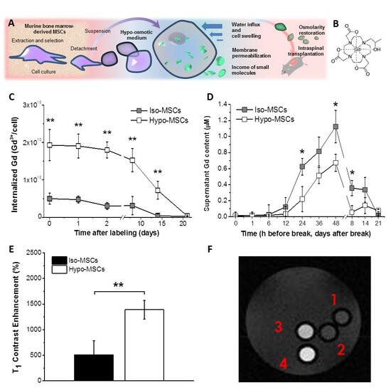
In this study, murine Mesenchymal Stem Cells (MSCs) labeled with the clinically approved MRI agent Gadoteridol through a procedure based on the hypo-osmotic shock were successfully tracked in vivo in a murine model of Spinal Cord Injury (SCI). With respect to iso-osmotic incubations, the hypo-osmotic labeling significantly increased the Gd 3 + cellular uptake, and enhanced both the longitudinal relaxivity ( r 1 ) of the intracellular Gadoteridol and the Signal to Noise Ratio (SNR) measured on cell pellets, without altering the biological and functional profile of cells.
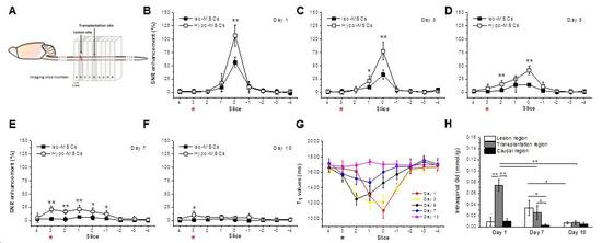
A substantial T 1 Contrast Enhancement after local transplantation of 3.0 × 10 5 labeled cells in SCI mice enabled to follow their migratory dynamics in vivo for about 10 days, and treated animals recovered from the motor impairment caused by the injury, indicating unaltered therapeutic efficacy.
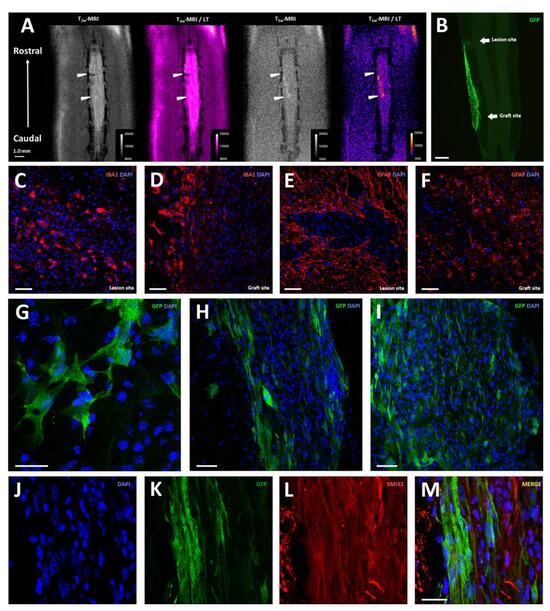
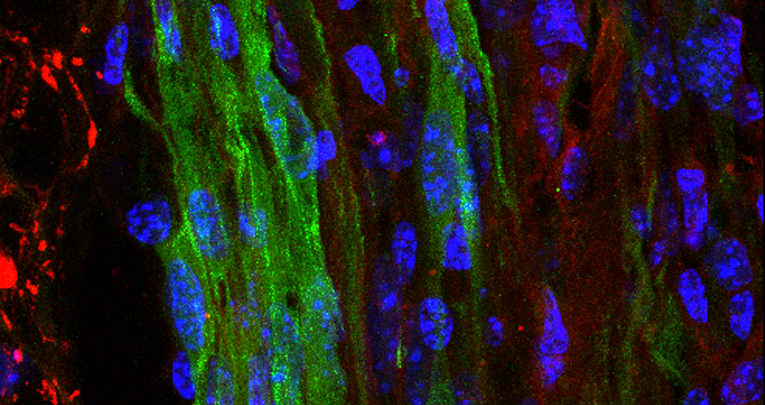
Finally, analytical and histological data corroborated the imaging results, highlighting the opportunity to perform a precise and reliable monitoring of the cell-based therapy.


