Stem Cell Reports
, 16 April 2020
Stem Cell-Derived Human Striatal Progenitors Innervate Striatal Targets and Alleviate Sensorimotor Deficit in a Rat Model of Huntington Disease
Dario Besusso # * 1 , Roberta Schellino# 2 , Marina Boido 2 , Sara Belloli 3 , Roberta Parolisi 2 , Paola Conforti 1 , Andrea Faedo 1 , Manuel Cernigoj 1 , Ilaria Campus 1 , Angela Laporta 1 , Vittoria Dickinson Bocchi 1 , Valentina Murtaj 3 , Malin Parmar 4 , Paolo Spaiardi 5 , Francesca Talpo 5 , Claudia Maniezzi 5 , Mauro Giuseppe Toselli 5 , Gerardo Biella 5 , Rosa Maria Moresco 3 , Alessandro Vercelli 2 , Annalisa Buffo 2* , Elena Cattaneo 1* .
*corresponding authors; #co-first authors.
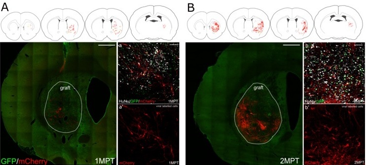
Huntington disease (HD) is an inherited late-onset neurological disorder characterized by progressive neuronal loss and disruption of cortical and basal ganglia circuits. Cell replacement using human embryonic stem cells may offer the opportunity to repair the damaged circuits and significantly ameliorate disease conditions. Here, we showed that in-vitro -differentiated human striatal progenitors undergo maturation and integrate into host circuits upon intra-striatal transplantation in a rat model of HD.
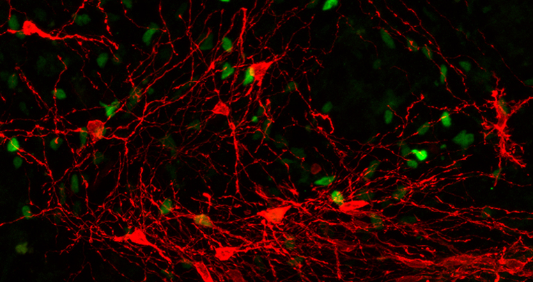
By combining graft-specific immunohistochemistry, rabies virus-mediated synaptic tracing, and ex vivo electrophysiology, we showed that grafts can extend projections to the appropriate target structures, including the globus pallidus, the subthalamic nucleus, and the substantia nigra, and receive synaptic contact from both host and graft cells with 6.6 ± 1.6 inputs cell per transplanted neuron. We have also shown that transplants elicited a significant improvement in sensory-motor tasks up to 2 months post-transplant further supporting the therapeutic potential of this approach.


