International Journal of Molecular Science
, 18 April 2020
TNFAIP3 Deficiency Affects Monocytes, Monocytes-Derived Cells and Microglia in Mice
Francesca Montarolo 1,2,3* , Simona Perga 1,2,4 , Carlotta Tessarolo 1,2 , Michela Spadaro 1,2 , Serena Martire 1,2 and Antonio Bertolotto 1,2
Abstract
The intracellular-ubiquitin-ending-enzyme tumor necrosis factor alpha-induced protein 3 (TNFAIP3) is a potent inhibitor of the pro-inflammatory nuclear factor kappa-light-chain-enhancer of activated B cell (NF-kB) pathway. Single nucleotide polymorphisms in TNFAIP3 locus have been associated to autoimmune inflammatory disorders, including Multiple Sclerosis (MS).
Previously, we reported a TNFAIP3 down-regulated gene expression level in blood and specifically in monocytes obtained from treatment-naïve MS patients compared to healthy controls (HC). Myeloid cells exert a key role in the pathogenesis of MS.Here we evaluated the effect of specific TNFAIP3 deficiency in myeloid cells including monocytes, monocyte-derived cells (M-MDC) and microglia analyzing lymphoid organs and microglia of mice.
TNFAIP3 deletion is induced using conditional knock-out mice for myeloid lineage. Flow-cytometry and histological procedures were applied to assess the immune cell populations of spleen, lymph nodes and bone marrow and microglial cell density in the central nervous system (CNS), respectively.We found that TNFAIP3 deletion in myeloid cells induces a reduction in body weight, a decrease in the number of M-MDC and of common monocyte and granulocyte precursor cells (CMGPs).
We also reported that the lack of TNFAIP3 in myeloid cells induces an increase in microglial cell density. The results suggest that TNFAIP3 in myeloid cells critically controls the development of M-MDC in lymphoid organ and of microglia in the CNS.
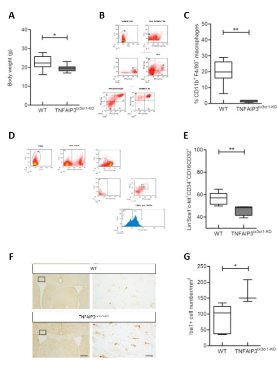
A - The myeloid specific TNFAIP3 deficient mice (TNFAIP3
cx3cr1-KO
) weigh less than control mice (WT).
B - Flow cytometry gating strategy used to evaluate myeloid cells.
C - The myeloid specific TNFAIP3 deficient mice (TNFAIP3
cx3cr1-KO
) show a reduction in the number of macrophages in comparison to control mice(WT).
D - Flow cytometry gating strategy used to evaluate the precursors of myeloid cells.
E - The myeloid specific TNFAIP3 deficient mice (TNFAIP3
cx3cr1-KO
) show a reduction in the number of precursors of myeloid cells in comparison to control mice(WT).
F - Representative histological images of the corpus callosum of the myeloid specific TNFAIP3 deficient mice (TNFAIP3
cx3cr1-KO
) and control mice (WT)used for the quantification of microglial cells density. Calibration bar 250 µm and 25µm.
G - The myeloid specific TNFAIP3 deficient mice (TNFAIP3
cx3cr1-KO
) show an increase in the microglia cell density in comparison to control mice(WT).
1
Neuroscience Institute Cavalieri Ottolenghi (NICO), Orbassano, 10043 Turin, Italy
2
Neurobiology Unit, Neurology–CReSM (Regional Referring Center of Multiple Sclerosis), AOU San Luigi Gonzaga, Orbassano, 10043 Turin, Italy
3
Department of Molecular Biotechnology and Health Sciences, University of Turin, 10126 Turin, Italy
4
Department of Neuroscience “Rita Levi Montalcini”, University of Turin, 10125 Turin, Italy


