


Wednesday 6
/12/23 h. 2:00 pm - Hybrid Seminar
John Mitrofanis, Scientific Director, Fonds Clinatec, Université Grenoble Alpes, France
Institute of Ophthalmology, University College London, United Kingdom
Lights-on for neurodegenerative disease: exploring the benefits of photobiomodulation
This seminar takes you on a journey, tracing the history of a somewhat serendipitous finding in the laboratory, to the translation of this finding to the clinic and its use on patients. The journey starts with a discussion over a cup of coffee between two old friends. They devised an experiment using a photobiomodulation device, one that delivered red to near-infrared light, on a few spare parkinsonian mice left over from other experiments. The thinking was that because photobiomodulation stimulates mitochondrial function, it may improve the mitochondrial dysfunction and protect neurones against the neurodegenerative insult (ie parkinsonian). After a week or so, it turned out that these photobiomodulation-treated mice had more surviving neurones than those that were not treated; in addition, they were found to have improved locomotor behaviour. This led to explorations in non-human primates, the gold-standard of all animal models of the disease. Here, in this species, the same beneficial outcomes were found, namely, less pathology and improved clinical signs. These experimental findings led to clinical interest and, as it stand now, clinical trials are underway testing the efficacy of several photobiomoduation approaches in patients. There are also encouraging, early indications that photobiomodulation is effective in Alzheimer's disease, with both neuroprotective and positive cognitive behavioural outcomes being evident in mouse models of the disease. We are in the process of starting a new series of studies that test the efficacy of photobiomodulation in Alzheimer's disease further, in both animal models and in patients.
Host: Corrado Calì
Monday 4
/12/23 h. 2:00 pm - Hybrid Seminar
Fernando De Castro, Cajal Institute, Spanish National Research Council, Madrid, Spain
When Cajal and the Spanish Neurological School showed the World how to study the brain
Santiago Ramón y Cajal (1852-1934) was still young and brimming with vital energy when he was tought in the reazione nera (1887), discovered years before by the Italian histologist Camillo Golgi in 1873. Cajal became absolutely passionated about the fine structure of the nervous system and started one of the last true epics of our modern history: the identification of nervous cells and their organisation to form the brain. His discoveries paving the birth of modern Neuroscience (the individuality of neurons/neuron theory, the synapses and the dendritic spines, the dynamic polarization of the neuron, the growth cones and the chemotactic hypothesis on their navigation…) saw the light while he was full professor at Barcelona (1887-1892): all this work was done by Cajal himself, alone, using his own private money to equip his laboratory… at his own home. Reticularists started to be defeated in 1889, but they were too powerful and stubborn to easily surrender the field to Cajal and the growing number of neuronists. Clairvoyant about the dimension of the scientific feat that he decided to face, as soon as he received international recognition and the Spanish authorities realized, Cajal received a modern laboratory in Madrid and started recruiting a handful of brilliant pupils who also contributed with significant and sometimes decisive discoveries to lay the foundations of modern Neuroscience and Neurology. Among many others, the most distinguished were Tello, Achúcarro, Pío del Río-Hortega, de Castro and Lorente de Nó, who worked side by side with El Maestro: the stud of their discoveries represents one of the zeniths of the collective discoveries ever in the History of Science. Their contributions range from the discovery of two of the three main types of glial cells to the description of the reverberant circuits that paved the way to Cybernetics, passing through the first identification of arterial chemoreceptors and the classification of nervous tumors. All together, they are known as the Spanish Neurological School, the School of Madrid or, directly, the School of Cajal. In words of Sherrington: “Never has anyone stated out on a great research more single-handed than at his beginning did Cajal. But as the years went by, if ever scientist had a school it was Cajal”.
Host: Alessandro Vercelli
Thursday 30
/11/23 h. 2:00 pm - Hybrid Seminar
Elif Keshinoz, Department of Anatomy, School of Medicine, Acibadem Mehmet Ali Aydinlar University - Istanbul, Turkey.
Mitochondrial alterations in Alzheimer’s Disease: Insights from the 3xTg Model
Aim: The 3xTg mouse model mimics key features of Alzheimer's disease (AD) through APP, PS1, and tau mutations. Mitochondrial dysfunction in AD results from interactions between these genes, leading to Aβ and tau deposits, damaging mitochondria, generating ER stress, and resulting in decreased ATP production, impaired neurons, and cell death. Mitochondrial dysfunction also leads to oxidative stress, inflammation, and the formation of amyloid plaques and neurofibrillary tangles, contributing to AD pathology. Mitochondria-ER contact sites (MERCs) are essential for cellular functions, including calcium signaling, lipid metabolism, and molecule exchange across organelles. An alteration may impact cellular calcium regulation, mitochondrial function, and Alzheimer's pathogenesis.
The study aims to investigate the role of mitochondrial alteration and MERCs in the 3xTg mice model of AD.
Materials and Methods: In this study, electron microscopic images of brain tissues from the CA3 regions of the hippocampus were taken at 10,000X magnification in 3xTg and Wild Type mice at 3, 8 and 12 months of age. Learning and memory deficits emerged at 3 months in the 3xTg mouse model, while cellular damage detectable through light microscopy became evident after 12 months. Various parameters were assessed to understand mitochondrial dynamics and morphology alterations. The images identified changes in mitochondrial architecture, such as mitochondrial number, area, and average widths. Additionally, the distance between mitochondria-endoplasmic reticulum contact sites (MERCs) was measured.
Results: Biophysical changes observed in mitochondrial architecture and MERCs shed light on the spatial organization and interactions between mitochondria and the endoplasmic reticulum. This biophysical analysis provides key clues for understanding changes in calcium signaling and cellular communication that affect mitochondrial dynamics and morphology in the CA3 region.
Conclusion:Understanding the complicated link between mitochondrial structure and MERCs in the 3xTg model could aid in the treatment of Alzheimer's disease via therapies aimed at protecting mitochondrial function.
Host: Stefania Raimondo
Wednesday 8
/11/23 h. 2:00 pm - Hybrid Seminar
Fernando De Castro, Cajal Institute, Spanish National Research Council, Madrid, Spain
Spontaneous remyelination: hot challenge for Multiple Sclerosis and the gate to go beyond
Spontaneous remyelination has been underestimated to date when treating patients with multiple sclerosis. The first results of a clinical trial that opened the door to this major challenge for Neurology were published around this time, 6 years ago. However, many attempts have failed to go further, basically due to weaknesses in the design of preclinical studies that have fueled false hopes. Our group has specialized in improving these preclinical studies and in exploring new compounds that can be incorporated in the future into the therapeutic arsenal with which to treat multiple sclerosis in combination with immunomodulators available in the clinic. We will show some of these cellular and molecular mechanisms, with special focus on small molecules and aptamers.
Host: Alessandro Vercelli
Wednesday 25
/10/23 h. 2:00 pm - Hybrid Seminar
Ana C. Cristóvão, CICS-UBI Health Sciences Research Centre, University of Beira Interior; NeuroSoV-Fastprinciple-Lda, UBIMedical - Covilhã, Portugal
Ionic-liquid Nox1 inhibitor to be used as a therapeutic solution to delay the progression of Parkinson's disease.
Parkinson's Disease (PD) is a chronic neurodegenerative disorder affecting up to 10 million people worldwide. Despite the efforts to develop a cure for PD, current therapeutic approaches can only target the symptoms. As symptomatic therapies lose effectiveness over time, patients end up with no therapeutic options. Therefore, delaying the disease progression became a promising solution to deal with this disorder.
PD pathogenesis is highly influenced by oxidative stress, and we have previously shown that ROS generated by NADPH oxidase 1 (Nox1) has detrimental impacts on dopaminergic neurons, being a valuable target for therapeutic developments. Inline, we have tested a chemical inhibitor ionic liquid for Nox1 inhibitor (N1inh-IL) capable of preventing neurodegeneration in experimental PD models.
In vitro studies showed that N1inh-IL has no cytotoxic effect on N27 neurons, while it significantly prevents the neurotoxic effect of two specific neurotoxins 6-hydroxydopamine (6OHDA) and 1-methyl-4-phenylpyridinium (MPP+). In vivo, the dopaminergic neuroprotective capacity of N1inh-IL, was evaluated in two animal models of PD, one induced by intrastriatal injection of 6OHDA and the other by chronic exposure to paraquat (PQ). The infusion of the N1inh-IL into the right ventricle did not cause neuronal toxicity, while it could prevent 6OHDA-induced neurodegeneration in the substantia nigra (SN) of mice. Moreover, four weeks after been exposed to PQ, the brain intraventricle diffusion or the intranasal delivery of N1inh-IL in rats was capable to prevent the motor dysfunction induced by the toxin and the accumulation of alpha-synuclein in the SN.
These results highlight that N1inh-IL can be an innovative therapy to reduce the speed of the progression of PD.
Host: Marina Boido, Serena Bovetti, Serena Stanga
Friday 20
/10/23 h. 2:00 pm - Hybrid Seminar
Francesco Moneta, Preclinical Imaging Division - Bruker BioSpin
Preclinical MRI and PET solutions and applications in neuroscience
Magnetic resonance imaging (MRI) and positron emission tomography (PET) are the core of Bruker portfolio in the field of preclinical research on small rodents. It will be presented the state of the art solutions for preclinical MRI and PET and some examples of their applications in the neurological field
Host: Alessandro Vercelli
Friday 13
/10/23 h. 3:00 pm
- Webinar
Chrystian Junqueira Alves, Icahn School of Medicine at Mount Sinai - New York
Mechanoregulation of Neurogenesis and Cancer: an Ancient Molecule Controlling Stem Cell Fate and Motility
Plexins are known as axon guidance receptors. However, Plexins originated in unicellular organisms greater than 600 million years ago. Dr. Junqueira Alves’ research aims to understand the fundamental role of Plexins during neurogenesis and cancer migration. Using cerebral organoids, he found that Plexin-B2-deficient neuroprogenitors undergo spontaneous neurogenesis. In cancer cells, Plexin-B2 is critical to promote invasiveness. Currently, he is developing novel strategies to accelerate the differentiation of stem cells by nuclear mechanics.
He is also exploring the role of plasma membrane and nucleus mechanoregulation for cancer migration. His research has the potential to accelerate the generation of neurons for disease modeling and discover new mechanisms of cancer migration for drug development.
Host: Roberta Schellino
Thursday 12
/10/23 h. 2:00 pm - Hybrid Seminar
Takehiro G. Kusakabe, Nanako Okawa, and Ayana Maruo, Konan University, Kobe, Japan
Ascidians: a simple chordate model to study the nervous system
Takehiro G. Kusakabe, PhD
The central nervous system of proto-vertebrate ascidians: a simple but informative model of the vertebrate brain
Vertebrates have evolved the complex brain with sophisticated cranial sensory organs and neuroendocrine systems. Invertebrate chordate ascidiansare the closest living relatives of vertebrates; the clade consisting of tunicates and vertebrates is called Olfactores.By using the ascidian Ciona intestinalis type A(also called Cionarobusta), we have revealed that ascidians have photoreceptive and gonadotropin-releasing hormone systems, which are similar to those of vertebrates. In this seminar, we introduce conserved and unique features of the ascidian nervous systemand present our recent findingsand evolutionary perspectives.
Nanako Okawa, PhD
Studies on neuron-glia interactions using optogenetics and Ca2+ imaging in Ciona larvae
The Ciona larva has a central nervous system homologous to that of vertebrates. Its nerve cord, the spinal cord homolog, is mainly composed of glial ependymal cells, along which run axons of cholinergic neurons that reside in the motor ganglion, which is the hind brain homolog. We visualized the activity of glial cells in the nerve cord of Ciona larvae by the Ca2+imaging method using G-CaMP8, and we found that active Ca2+ transients were associated with tail motions and swimming behavior in response to light. We further analyzed the relationship between neuronal activation and glial cell activity using optogenetics in combination with Ca2+ imaging. Our resultsrevealed that the glial ependymal cells in the nerve cord receive cholinergic input from the motor ganglion. Consistently, receptors for various neurotransmitters were shown to be expressed in the glial cells of the nerve cord. Our findings suggest that the glial cells actively interact with neurons and are involved in the control of swimming behavior.
Ayana Maruo, MSc
Spatial transcriptomic analysis of the adult brain of Ciona intestinalis type A
The swimming larvae of ascidians metamorphose into sessile adults, and the adult brain is formed after metamorphosis from a part of the larval brain. In contrast to our detailed knowledge of the larval brain, little is known about the function, cellular composition, and developmental mechanisms of the adult brain. To lay the foundation for elucidating the structure and functions of the adult ascidian brain and its developmental mechanisms, we performed spatial transcriptome analysis of the adult brain of the ascidian Ciona intestinalis type A. Our analysispresents, for the first time, comprehensive gene expression data of the ascidian adult brain and gives insights into its structure and function.
Host: Giovanna Gambarotta
Friday 22
/9/23 h. 2:00 pm - Hybrid Seminar
Pascal Hot, Université Mont-Blanc - Chambery
Functional specialisation of the medial temporal lobe and hippocampus: the representational approach
A growing body of research has shown that medial temporal regions play a role outside the memory domain, as they would be specialised in processing certain types of representations. In this view, the hippocampus and perirhinal cortex (PRC) would represent scenes and entities, respectively, independently of the memory or non-memory operation performed on it (Cowell et al., 2019). Using both fMRI (Gardette et al., 2023a, 2023b) and patients with right or left temporal lobectomy, we tested this prediction in the operations of pattern-completion (i.e., the operation involved in recollection), familiarity-based recognition and rejection, and visual discrimination of scene and object images.
We will present our recent data from these studies supporting the representational view of the MTL: the engagement of these regions in processes such as recollection and familiarity would be determined by the representation involved rather than by the operation.
Host: Serena Bovetti
Friday 8
/9/23 h. 3:00 pm - Webinar
Introduction to BioRender
Host: Letizia Marvaldi
Friday 7
/7/23 h. 2:00 pm - Hybrid Seminar
Mike Fainzilber, Weizmann Institute of Science, Israel
Neuronal Injury Signaling: SINEs of Growth?
Importins, molecular motors and RNA binding proteins function in a bidirectional mechanism of intracellular communication, consisting of anterograde RNA transport, local translation at axon tips, and retrograde transport of the resulting proteins, for neuron length sensing and growth control. The talk will focus on recent findings revealing an unexpected role for a specific subgroup of non-coding RNAs in this mechanism.
Host: Letizia Marvaldi
Wednesday
28/6/23 h. 2:00 pm - Hybrid Seminar
Hakeem O. Lawal
Delaware Center for Neuroscience Research and Department of Biological Sciences, State University, Dover - Delaware
Drosophila models of Parkinson’s Disease and Aging
Drosophila is an excellent model system for the study of many human neurological disorders and states. Parkinson’s disease represents one such example. The second most common neurological disease, it is characterized by the loss of dopaminergic neurons of the substantia nigra pars compacta. It has no known cure and current treatments cause severe side effects.
This status quo necessitates the deployment of every model system available to help accelerate progress towards understanding both the cause of the disease and the development of viable treatment strategies.
Here we present findings from our lab that demonstrate the utility of Drosophila as a model system to understand the possible underlying causes of this disease and to develop effective treatment strategies. Similarly, we present work from group establishing a role for cholinergic synaptic transmission in the central nervous system on behavioral changes that occur during aging. Moreover, we show that overexpression of the vesicular acetylcholine transporter (VAChT), which mediates the packaging of acetylcholine into synaptic vesicles for exocytotic release, causes a reduction in lifespan and a decline in ACh-linked behaviors during aging. Together, our work adds importantly new light to the contributions of Drosophila as a model for advancing our understanding of both normal and pathological aging.
Host: Ferdinando Di Cunto
Friday
23/6/23 h. 2:00 pm - Webinar
Vanessa De Luca, IIT - Genova, Italia
GENDER DIMENSION IN RESEARCH
Horizon Europe is the new European Research & Innovation funding program, following Horizon 2020. Across its commitments, the gender dimension emerges as a cross-cutting objective. Particularly, a set of interventions promises to tackle gender inequalities by dismantling material and cultural barriers that leave women at a disadvantage within the research sector. This talk will focus on conceptual and practical issues concerning the gender dimension in research exploring a few practical examples of how to integrate gender-sensitive contents and methods in research, and apply them when developing new projects and/or funding procedures under the Horizon framework.
Host: Stefano Zucca
Thursday
15/6/23 h. 2:00 pm -
Hybrid Seminar
Shimon Ben Shaabat, Department of Biochemistry & Pharmacology, Faculty of Health Sciences, Ben Gurion University, Israel
Role of phytocannabinoids in neuroinflammation and Nanotechnological approach for topical-dermal delivery and intranasal-direct brain targeting
Multiple sclerosis (MS) is a widespread chronic neuroinflammatory and neurodegenerativeDisease. CBG, and CBDA,phytocannabinoids, have attracted significant pharmacological interest due to their non-psychotropic nature. We studied the effects of these compounds on microglial inflammation in vitro, followed by an in vivo study. CBG and CBDA attenuated the microglial production of NO in BV2 microglia and primary glial cells and reduced iNOS expression. TNF-a on the other hand was decreased by CBG but increased by CBDA. The same was found in MSin vivomodel, experimental autoimmune encephalomyelitis (EAE). The clinical scores of EAE mice were attenuated and lumbar sections of EAE mice showed enhanced neuronalloss.Despite the potential activity, the delivery of phytocannabinoidsto the periphery and to CNS remains challenging. We have developed a new particulate system capable of deliveringphytocannabinoidsinto the periphery by transdermal delivery and to the brain via the intranasal route. In cultures of LPS-induced inflamed BV2 cells, the phytocannabinoid-loaded starch nanoparticles demonstrated low toxicity while effectively reducedNO production and IL-6 levels. Intranasal administration of CBD-loaded starch nanoparticles resulted in higher levels of CBD in the brain than an identically administered CBD solution.
Host: Ilaria Bertocchi and Carola Eva
Friday 9/6/23 h. 2:00 pm -
Hybrid Seminar
Marta Valenza, Department of Biosciences, University of Milan
Astrocyte-neuron interplay in the cholesterol dysfunction in Huntington’s disease brain: from the mechanism to therapeutics
In the adult brain, neurons require local cholesterol production, which is supplied by astrocytes. Cholesterol biosynthesis is severely reduced in the brain of Huntington’s disease (HD), a genetic adult-onset neurodegenerative disease characterized by synaptic dysfunction and motor and cognitive defects. The defect occurs inastrocytes with detrimental consequences on HD neurons’ activities. The underlying molecular mechanism is a reduced activity of SREBP2, the transcription factor that activates the expression of many genes involved in cholesterol synthesis.
In the last years, different in vivo strategies were developed to counteract cholesterol dysfunction in HD mouse models either supplying exogenous cholesterol with brain-permeable nanoparticles or enhancing endogenous cholesterol within the HD brain. I will present an overview of thesecholesterol-based strategies and their translational potential. Then, I will focus on the gene therapy approach to force endogenous cholesterol biosynthesis in the striatal astrocytes of HD miceto highlight the relevance of the astrocyte-neuron interplay in HD pathogenesis in vivo.
Host: Marina Boido
Tuesday 6/6/23 h. 2:00 pm -
Hybrid Seminar
Marco Cambiaghi, University of Verona - Dep. of Neurosciences, Biomedicine and Movement Sciences
Electrified brains: from the torpedo fish to transcranial direct current stimulation
The idea of modulating brain activity with a non-invasive approach has always been one of the major goals of neurophysiology and to a broad extent, of modern neurology and psychiatry, since the associated disorders are often the consequences of dynamic plastic changes of the neural networks. Well before the discovery of the physical phenomenon, electricity was used as a therapeutical tool but only the last few decades saw the development of effective non-invasive neuromodulatory techniques. Among them, transcranial direct current stimulation (tDCS) has recently emerged as a safe and economic tool to guide neuroplasticity and modulate cortical function by tonic stimulation with weak direct currents. Despite its wide use in human studies, some underlying mechanisms of action have been clarified only recently and the vast majority is still to be elucidated. In recent years, we are focusing on the study of indirect effects of tDCS and the influence of brain state during stimulation, in different preclinical models in both physiological and pathological conditions. In particular, we explored prefrontal tDCS influence on dorsal raphe activity and, in the motor cortex, the effects of combining tDCS with physical activity.
Host: Enrica Boda
Friday 19/5/23 h. 2:00 pm
- Hybrid Seminar
Silvia Gancheva Marinova e Antoaneta Georgieva (University of Varna, Bulgaria)
Silvia Gancheva Marinova, MD, PhD
Pharmacologically induced changes in osteocalcin levels – metabolic and central nervous system effects in healthy and metabolic rats
Osteocalcin is a bone-derived protein involved in the regulation of energy metabolism and CNS functions in rodents. Its serum concentrations can be modified pharmacologically in opposite directions through administration ofvitamin K antagonists and bisphosphonates. The current presentation describes the resulting changes in energy metabolism and brain functions.
Antoaneta Georgieva, MD, PhD
Effects of Aroniamelanocarpa fruit juice and its component chlorogenic acid on the ovariectomy-induced behavioral changes in rats
Presentation of our research on the behavioral changes in a rat model of ovariectomy-induced estrogen deficit and the effects of a 10-week treatment with Aroniamelanocarpafruit juice in two different doses or chlorogenic acid.
Host: Ilaria Bertocchi/Carola Eva
Friday 21/4/23 h. 2:00 pm
- Hybrid Seminar
Alessandro Ferrarini, Account Manager Starlab
The Sustainable Laboratory
It is universally acknowledged that the Pharmaceutical Industry and Scientific Research sector are highly polluting, in terms of CO2 emissions, plastic waste and water usage. A study conducted in2015 estimated that labs worldwide consume around 5 million tons of plastic and that a research lab requires 5 to 10 times the amount of energy used in an office of the same size.
The current situation is alarming and rapidly deteriorating. The Pharma and Life Science Sector has one of the largest carbon footprints globally, estimated to be even higher than the automotive industry. While the Pharmaceutical and healthcare sectors are clearly the biggest contributors, scientific research also play a role in the overall result, with plastic waste and energy consumption being the most significant factors.
Starlab mission is to continually look for intelligent, climate-friendly products and processes. We try to raise awareness among scientists and present possible solutions: The new TipONE generation saves up to 40% in plastic and the smart gloves packaging optimizes space and transportation. Starlab is constantly thinking about sustainability, with the ultimate goal to become a Green Company in every aspect. So, Let’s get green Together.
Host: Serena Stanga
Friday 7/4/23 h. 2:00 pm
- Webinar
Alain Prochiantz, Emeritus Professor at College de France and Chief Scientific Officer BrainEver SAS
OTX2 and EN1 homeoprotein transduction, from physiopathology to therapeutic strategies
Intercellular transfer has now been demonstrated for 150 homeoprotein transcription factors. However, the physiological functions served by this novel signaling pathways have only been studied for a handful of them, including OTX2 and ENGRAILED. The conference will focus on the role of OTX2 and ENGRAILED-1 signaling in the regulation of cerebral cortex plasticity and spinal cord a-Motoneuron physiology, respectively. The consequences of the latter recent findings in the development of original therapeutic strategies in mood disorders and Amyotrophic Lateral Sclerosis will be discussed.
Host: Serena Stanga
Friday 24/3/23 h. 2:00 pm
- Hybrid Seminar
Fiorenza Stagni, Università degli Studi di Bologna
Potential of early pharmacotherapies for the improvement of intellectual disability in Down syndrome: lesson from the Ts65Dn mouse model
Down syndrome (DS) is a relatively high-incidence genetic condition caused by the triplication of chromosome 21. Gene triplication may compromise different body functions but intellectual disability represents the unavoidable hallmark and the most invalidating aspect of this pathology. Intellectual disability is mainly attributable to neurogenesis and dendritogenesis alterations that can be traced back to fetal life stages. Although the progressive improvement in medical care has led to a notable increase in life expectancy for people with DS, there are currently no effective therapies for intellectual disability in DS. Since neurodevelopmental defects are present starting from fetal life stages, early pharmacological interventions are likely to represent a good strategy to improve brain development in DS. With this idea in mind, our research group has examined the efficacy of different pharmacotherapies administered during the prenatal or early postnatal period in the Ts65Dn mouse, a model that recapitulates many anatomical and functional alterations of DS.This talk will describe and discuss the most suitable time windows for treatment and some of the attempted pharmacotherapies targeted to pathways that are known to be deranged in DS, that have proved to be effective in restoring trisomy-linked neurodevelopmental defects and cognitive performance in the Ts65Dn mouse model.In view of the good translational impact of some of these tested therapies, our preclinical findings may open the way for clinical trials in individuals with DS, thereby improving their life conditions.
Host: Francesca Montarolo
Wednesday 22/3/23 h. 2:00 pm
- Hybrid Seminar
Christel Genoud, Electron Microscopy Facility, University of Lausanne
The benefits and challenges offered by visualization of large volume with light and scanning Electron Microscopy
In recent years, the field of microscopy has advanced significantly, allowing for the visualization of larger biological structures in 3 dimensions at ever-increasing resolutions. In cellular electron microscopy, this evolution is allowed by the technological developments in volume EM and Correlative multi-modal imaging. These techniques offer a wealth of benefits, including the ability to visualize large volumes of tissue with remarkable detail as well as target rare events in large volumes. However, there are also significant challenges associated with these techniques, including sample preparation, data acquisition, and image processing.
In this talk, we will explore some of the benefits and challenges of these approaches, with a focus on their applications in biological research. We will discuss recent advances in these techniques, including the use of machine learning to aid in data analysis, and highlight some projects that have been made using these methods. Ultimately, we will demonstrate that while these techniques are not without their challenges, they offer tremendous potential for advancing our understanding of the biological world.
Host: Corrado Calì
Friday 17/3/23 h. 2:00 pm
- Webinar
Elena Choleris, Department of Psychology and Neuroscience Program, University of Guelph, Ontario, Canada
Neuroendocrinology of social cognition in female and male mice
Recent findings demonstrate rapid hormonal facilitation of social cognition in various brain regions of female a male mice. In females, in the Dorsal Hippocampus estradiol’s rapid facilitating effects are broad across multiple (but not all tested) learning tasks. In the Paraventricular Nucleus of the Hypothalamus (PVN), Medial Amygdala (MeA) and Medial Prefrontal Cortex, instead, the effects appear specific to social cognition, other types of learning tasks being unaffected. In the PVN and MeA those effects depend upon the action of MeA oxytocin receptors and do not extend to males. In males, in the Bed Nucleus of the Stria Terminalis, estradiol, testosterone and dihydrotestosterone all rapidly facilitate social recognition while inversely affecting object recognition, and those effects depend upon Arginine Vasopressin receptors V1aR in the Lateral Septum. Together, these investigations are highlighting the rapid hormonal regulation of brain networks of females and males subserving social cognition, further advancing our understanding of hormonal regulation of the social brain.
Host: Gruppo Panzica Gotti
Thursday 9/3/23 h. 2:00 pm
-
Hybrid seminar
Philip Greulich, University of Southampton
Mathematical modelling in cell biology: why and when is it useful?
Mathematical and computational modelling has become an increasingly popular tool in the biological sciences. Yet, often modelling is being employed without gaining much value from it, or in ways where machine learning approaches would be superior. Sadly, wrong use of model fitting can also lead to plainly wrong results. In this talk, I wish to outline some contemporary uses of mathematical modelling in (cell) biology and explain which modelling approaches are generally fruitful and can enrich biological research, in ways that are not achievable by plain experimental approaches.
I will exemplify this on some research projects about clonal dynamics in developing tissue -- mouse mammary gland during pregnancy and microglia in the developing brain -- where experimental data and mathematical modelling were successfully combined to drive the discovery of stem cell fate choice patterns. These examples will show where the pitfalls of mathematical modelling lie and how to circumnavigate them to achieve scientifically sound outcomes that go beyond the reaches of experimental research.
Host: Federico Luzzati
Friday 10/3/23 h. 2:00 pm
- Hybrid seminar
Paolo Giacobini, INSERM Research Director, Lille Neuroscience & Cognition
Development and Plasticity of the Neuroendocrine Brain
The scent-sational role of GnRH neurons
Gonadotropin-releasing hormones (GnRH) neurons are the master regulators of fertility in vertebrates. Hypothalamic GnRH-secreting neurons release their hormone through the median eminence (ME) into the hypophyseal portal system to stimulate the production and release of pituitary gonadotropins, which regulate the development and function of the gonads. To ensure reproductive success, GnRH neurons have to process and integrate various internal and external cues to elicit the most adapted neuroendocrine responses.
We recently identified, both in mice and humans, a sub-population of extra-hypothalamic GnRH neurons located in the olfactory bulb (OB), whose role has never been investigated. Combining mouse genetics with Cre-dependent viral tracing approaches, and 3D-imaging of whole-mouse heads, we revealed that OB-GnRH neurons project neurites contacting the vomeronasal organ, the chemosensory system that perceives and processes stimuli related to social and reproductive behaviors in many species of vertebrates. In addition, OB-GnRH neurons send long projections to the hypothalamic areas involved in the control of gonadotropin release.
Bidirectional chemogenetic neuromodulation combined with behavioral testing, electrophysiological recording and two-photon in vivocalcium-imaging, demonstrated a novel role for this extra-hypothalamic GnRH neuronal population as a central regulatory hub linking pheromonal stimulation with the neuroendocrine response regulating reproduction and mating behavior.
Host: Silvia De Marchis
Friday 3/3/23 h. 2:00 pm
- Webinar
Wenhui Huang, Universität des Saarlandes Homburg, Germany
Adenosine control of glial fate and functions
Extracellular adenosine is mainly formed from the sequential ATP metabolism by a series of hydrolases, in which ecto-5’-nucleotidase (Nt5e, also known as CD73) performs the last step converting AMP to adenosine. Under cellular stress conditions, such as inflammation, hypoxia, etc., extracellular adenosine can be drastically increased. In the CNS, adenosine serves as a neuromodulator by triggering various G-protein-coupled adenosine receptors (ARs) of the A1, A2a, A2b and A3 subtypes among which A1 receptors (A1ARs), coupled to Gi/o proteins, are the most abundant subtype. A1ARs are widely expressed in the brain, including neurons, microglia, astrocytes, and oligodendrocyte (OL) lineage cells.
Transcriptomic studies revealed the highest A1AR expression levels in OL lineage cells and astrocytes, indicating an important role of adenosine signaling in regulating the fate and functions of these glial cells. In my talk, I will introduce our ongoing work studying the in vivo functions of adenosine signaling in glial cells by analysing cell type-specific A1AR deficient mice in a cuprizone-induced de-/remyelination model as well as in a peripheral lipopolysaccharide (LPS) injection model.
Host: Enrica Boda
Friday 10/2/23 h. 2:00 pm
- Hybrid seminar
Samuele Negro, Department of Biomedical Sciences, University of Padova IT
CXCR4: a new target to boost peripheral nerve regeneration
The peripheral nervous system (PNS) is the part of the nervous system outside the brain and spinal cord that relays signals between the central nervous system (CNS) and the rest of the body. It is composed mainly of: 1) neuronal cells, in particular a combination of motor, sensory and autonomic neurons; 2) Schwann cells (SCs), glial cells which ensheaths nerves in a layer of myelin and provide trophic support; 3) other non-neuronal cells such as fibroblast and satellite cells. Despite the PNS has an intrinsic ability for repair and regeneration to a certain extent, differently from the CNS, peripheral nerve injuries represent an important clinical issue with insufficient or unsatisfactory therapeutic approaches.
The process of nerve regeneration is complex, involving many factors concerning to the neuron and the cellular environment, and is still far from being understood. Our research group has contributed to shedding light on some of the mechanisms that govern PNS regeneration. In particular, we have recently discovered that the molecular axis orchestrated by the chemokine CXCL12α and its receptor CXCR4 is a novel signalling pathway that powerfully stimulates peripheral nerve regeneration.
Host: Roberta Schellino
Friday 27/1/23 h. 2:00 pm
- Hybrid seminar
Indrek Koppel, Tallinn University of Technology, Estonia
Cell type-specific omics of neurons and glia
For studying transcriptomes in a cell type-specific manner researchers can choose between single-cell strategies and affinity purification of ribosome-associated mRNA. Single cell proteomics is in very early stages of development, but cell-specific labeling and affinity purification of proteins has been achieved using genetic tools, ensuring incorporation of affinity tags in cell types of interest. In this talk, I will discuss currently available tools in cell type-specific proteomics and introduce a method that leverages a puromycin inactivating enzyme for achieving cell type specificity. I will describe the use of this strategy for studying protein synthesis in co-cultures of rat cortex cells. In addition, I will talk about our efforts in studying neuron-astrocyte communication using tools for cell-specific activation and translatomics.
Host: Letizia Marvali
Friday 20/1/23 h. 2:00 pm
- Hybrid seminar
Tommaso Pizzorusso, BIO@SNS laboratory, Scuola Normale Superiore of Pisa; Institute of Neuroscience CNR, Pisa
Genetic and environmental regulation of visual cortical plasticity
The visual cortex is characterized by developmental periods of high plasticity designated critical periods. However, environmental factors are able to modulate plasticity levels also in adult animals. Indeed, exposing animals to an enriched environment (EE) has dramatic effects on brain structure, function, and plasticity also in adult animals. The poorly known ‘‘EE-derived signals’’ mediating the EE effects are thought to be generated within the central nervous system. In the talk I will report data about intrinsic regulators of cortical plasticity and the interaction with signals originating from the periphery that can be changed by life style. The role of the gut microbiota and the effect of diet will be discussed
Host: Serena Bovetti
Agenda
Area Ricercatori
Guarda il video
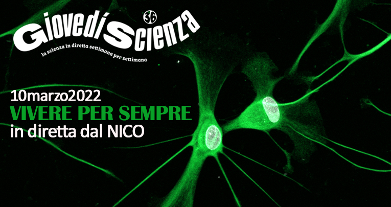
GiovedìScienza racconta la ricerca al NICO
Vivere per sempre.
Una popolazione sempre più longeva, i suoi problemi e le risposte della ricerca
Hai perso la diretta? Guarda ora il video di GiovedìScienza al NICO: una puntata in diretta dai nostri laboratori dedicata alla ricerca sull'invecchiamento.